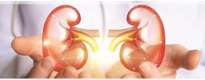
Kidney stone
Kidney Stone
UROLITHIASIS is the development of solid piece of mineral/material in the urinary tract. It maybe solitary or multiple, renal or ureteric depending on the number and location of calculus. Stones maybe primary or secondary based on their etiology, and it may vary in their composition and size. Most commonly stones are composed of CALCIUM OXALATE,while other components include uric acid, calcium/magnesium/ammonium phosphate,xanthine etc.
Most stones form due to a combination of genetics and environmental factors. RISK FACTORS include high urine calcium levels; obesity; excess of certain foods(spinach,chocolates,nuts,tomato etc); some medications, supplements; hyperparathyroidism; gout and not drinking enough fluids. Urolithiasis is a WIDELY PREVELANT disease, between 1% and 15% of people globally are affected by kidney stones at some point in their lives. In 2015, 22.1 million cases occurred, resulting in about 16,100 deaths.
The SYMPTOMS of renal stones include RENAL COLIC(an intermittent,excruciating pain which usually originates from the flank region of the concerned side) and radiates to the back or down to the groin region,depending on the location of the stone), nausea,vomiting,dysuria(especially in case of lower ureteric calculus), hematuria(blood in urine) among others.
The pain typically comes in waves, with each episode of colic lasting for 15-30 minutes. Presence of fever or burning micturition usually signifies super-added urinary tract infection and is usually considered a surgical emergency which would require immediate surgical intervention. Sometimes when the infection spreads to the blood(septicemia), patient may even present in shock, which require lengthy hospital stay, possible intensive care in an ICU setup and prolonged recovery,apart from general resuscitative measures including IV fluids, high grade antibiotics and inotropes (medicines to increase BP).
Patient can also present with reduced urine output or even renal failure, especially if there are stones on both sides or if the patient has a solitary functioning kidney. At presentation the patient is INVESTIGATED to find out the number and location of the stone(s), as well as additional information like presence/absence of super-added infection, renal function etc of the patient.
Apart from routine HEMATOLOGICAL work-up(CBC, KFT, Viral markers, Coagulation profile) and URINE TESTS (Urine R/M, Urine C/S), patient undergoes RADIOLOGICAL TESTS like Ultrasound and/or plain CT(Computed Tomography) of the KUB region. Patient may also be required to undergo Blood C/S if septicemia is suspected.
Once all the investigations are done and reviewed, decision regarding management is done. MANAGEMENT STRATEGIES include conservative or surgical interventions. When the size of the stone is 5 mm or less the chances of stone expulsion with the help of hydrotherapy along with alpha blockers(namely Tamsulosin/Silodosin/Alfuzosin) and painkillers, is quite high. This CONSERVATIVE line of management however cannot be tried in the presence of fever or intractable pain(not relieved by taking painkillers even at optimal doses).
In case of larger obstructive/symptomatic stones,operative management is mandated. Under certain circumstances(like Kidney stones <1cm, with favourable HU value on Plain CT Scan), ESWL (Stones are broken with external shock waves,delivered in a focussed and targetted manner over the stone) can be attempted.
Common modality of surgery is ENDOSCOPIC(through natural urinary tract) or minimally invasive(PCNL – through a small hole at the back – Size of which has undergone progressive miniaturisation – Mini-/Ultra-Mini/Micro-PCNL) or LAPAROSCOPIC or Open surgery. Endoscopic surgery(URS,RIRS) is done when stones are stuck in the ureter or kidney. In this form of surgery,the surgeon approaches the stone with the help of thin instruments inserted through the natural urinary tract of the patient, breaks it with the help of laser/lithotripter, and evacuates the stone fragments.
This way,the entire surgery is done without any single external cut or scar. A Double-J stent may or may not be placed,which is usually removed after 2 weeks under Local Anesthesia on a daycare admission basis. When the size of the renal stone exceeds 2 cm, or when RIRS is not feasible due to various reasons, then a patient is offered PCNL(Mini-/Ultra-Mini/Micro) , which requires a small hole to be made on the back,on the side of the kidney stone, and then endoscopic instruments are used to break the stone and remove it.
A DJ stent may or may not be placed and a small tube (PCN) may or may not be placed through the puncture site,which is removed after a day or two before discharge.
In case of larger stone being stuck in the ureter,or in case of failed endoscopic surgery or in case of paucity of endoscopic instruments, a Laparoscopic surgery maybe attempted. A small cut is made over the concerned segment of the ureter and the stone is extracted out and the cut is then repaired with the help of sutures over a Double-J stent which is removed after 2-3 weeks under Local Anesthesia. Open Nephro/ureterolithotomy is attempted if laparoscopy fails or is not feasible/available.
A general preventive method for all types of stones, is to drink lots of oral fluids especially water. While all surgical modalities require atleast 1-2 days of admission and administration of some form of anaesthesia (General/Regional), ESWL and hydrotherapy can be performed on OPD/Daycare basis.
When the stone size is larger then single stage clearance may not be possible, and may require a staged approach. The stone fragments are sent for analysis , based on which certain dietary modifications are suggested to prevent recurrence.
Dr Karan r rawat safe surgery center is a renowned kidney stone doctor in Agra


